체외 충격파 쇄석술
체외 충격파 쇄석술(體外衝擊波碎石術, Extracorporeal shock wave lithotripsy, ESWL)은 신장결석과[1] 담석(쓸개나 간의 돌)의 무침습 치료 방법이며 음향 펄스를 이용한다. 타석[2]과 췌장석에도 사용되는 것으로 보고되었다.[3]
| 체외 충격파 쇄석술 | |
|---|---|
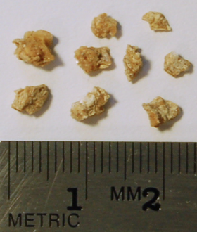
 1 cm의 옥살산칼슘 돌조각 중 일부가 쇄석술을 이용해 쇄석어 있다. | |
| ICD-9-CM | 98.5 |
| MeSH | D008096 |
역사
편집1969년을 기점으로 독일 국방부의 자금 지원을 받은 도니어(Dornier)는 조직에 대한 충격파의 영향에 대한 연구를 시작하였다. 1972년 도니어 메디컬 시스템즈(Dornier Medical Systems)가 수행한 기초 연구를 기반으로 하여 뮌헨 대학교의 비뇨기학 책임자 Egbert Schmiedt와 합의에 도달하였다. 도니어 쇄석술의 개발은 여러 원형들을 통해 발전하였으며, 궁극적으로는 1980년 2월 최초로 인간에 충격파 쇄석술(SWL)을 사용하여 치료하기에 이른다. 도니어 HM3 쇄석기의 개발과 배급은 1983년 말에 시작되었으며 충격파 쇄석술은 1984년 미국 식품의약국에 의해 승인되었다.[4]
비침습 치료
편집쇄석기는 외부에서 적용되고 고밀도로 집중되는 음향 펄스를 사용하여 부수적 위험을 최소화하면서 쇄석을 시도한다. 환자를 가만히 있도록 하고 잠재적인 불편함을 감소시키기 위해 환자에게 시술할 때 진정작용 또는 마취를 하는 것이 보통이다.[5] 환자는 기구의 침상에 눕고 등은 물로 채워진 기기에 지탱하며 콩팥이 있는 곳까지 위치시킨다. 형광 투시 엑스선 촬영 시스템이나 초음파 촬영 시스템을 사용하여 치료를 위해 돌 방향으로 조준하게 된다.
체외 충격파 쇄석술은 확고한 결석 치료를 위한 최소한의 침습을 동반하지만 RIRS(retrogade intrarenal surgery) 또는 경피적신쇄석술 또는 레이저 쇄석술을 동반한 요관경검사 등 기타 다른 침습 치료 방식들에 비해 더 낮은 결석 제거율을 보인다.[6] 돌 조각들이 빠져나가는 데까지는 수일에서 1주가 소요될 수 있으며 환자와 시술의 성공 정도에 따라 온화한 정도에서 극심한 통증을 일으킬 수 있다. 이 시기에 환자는 가능한 물을 많이 마실 것을 지시받는다.
체외 충격파 쇄석술 자체가 위험이 없는 것은 아니다. 충격파 그 자체 외에도 소변 매개체의 움직임에 의해 형성되는 진공현상의 거품들은 모세혈관 손상, 신실질, 피막하 출혈을 일으킬 수 있다. 이렇게 되면 신부전과 고혈압과 같은 장기간의 후유증을 일으킬 수 있다. ESWL의 전반적인 합병증 비율은 3%에서 20% 사이이다. 종종 환자들은 감염을 경험하므로 의료 전문의는 발열이 발생할 경우 조속히 의료적인 지원을 받을 것을 충고한다.
추가 문헌
편집- Abe T, Akakura K, Kawaguchi M, Ueda T, Ichikawa T, Ito H, 외. (2005). “Outcomes of shockwave lithotripsy for upper urinary-tract stones: a large-scale study at a single institution”. 《J. Endourol.》 19 (7): 768–73. doi:10.1089/end.2005.19.768.
- Albala DM, Assimos DG, Clayman RV, Denstedt JD, Grasso M, Gutierrez-Aceves J, Kahn RI, Leveillee RJ, Lingeman JE, Macaluso JN, Munch LC, Nakada SY, Newman RC, Pearle MS, Preminger GM, Teichman J, Woods JR (2001). “Lower pole I: a prospective randomized trial of extracorporeal shock wave lithotripsy and percutaneous nephrostolithotomy for lower pole nephrolithiasis-initial results”. 《The Journal of Urology》 166 (6): 2072–80. doi:10.1016/s0022-5347(05)65508-5.
- Anagnostou T, Tolley D (2004). “Management of ureteric stones”. 《Eur. Urol.》 45 (6): 714–21. doi:10.1016/j.eururo.2003.10.018. PMC 3180306.
- Auge BK, Preminger GM (2002). “Update on shock wave lithotripsy technology”. 《Curr. Opin. Urol.》 12 (4): 287–90. doi:10.1097/00042307-200207000-00005.
- Chacko J, Moore M, Sankey N, Chandhoke PS (2006). “Does a slower treatment rate impact the efficacy of extracorporeal shock wave lithotripsy for solitary kidney or ureteral stones?.”. 《J. Urol.》 175 (4): 1370–3. doi:10.1016/s0022-5347(05)00683-x.
- Chaussy CG, Fuchs GJ. "Current state and future developments of noninvasive treatment of human urinary stones with extracorporeal shock wave lithotripsy. J Urol. 1989;141(3 Pt 2):782-9.
- Collins JW, Keeley FX (2002). “Is there a role for prophylactic shock wave lithotripsy for asymptomatic calyceal stones?”. 《Curr. Opin. Urol.》 12 (4): 281–6. doi:10.1097/00042307-200207000-00004.
- Delius M (1988). “This month in Investigative Urology: effect of extracorporeal shock waves on the kidney”. 《J. Urol.》 140 (2): 390.
- Hayes J, Ding S (2012). “Pancreatic stone and treatment using ERCP and ESWL procedures: a case study and review”. 《NZMJ》 125 (1361).
- Joshi HB, Obadeyi OO, Rao PN (1999). “A comparative analysis of nephrostomy, JJ stent and urgent in situ extracorporeal shock wave lithotripsy for obstructing ureteric stones”. 《BJU Int.》 84 (3): 264–9. doi:10.1046/j.1464-410x.1999.00174.x.
- Kim FJ, Rice KR (2006). “Prediction of shockwave failure in patients with urinary tract stones”. 《Curr. Opin. Urol.》 16 (2): 88–92. doi:10.1097/01.mou.0000193373.22318.68.
- Krambeck AE, Gettman MT, Rohlinger AL, Lohse CM, Patterson DE, Segura JW (2006). “Diabetes mellitus and hypertension associated with shock wave lithotripsy of renal and proximal ureteral stones at 19 years of followup”. 《J. Urol.》 175 (5): 1742–7. doi:10.1016/s0022-5347(05)00989-4.
- Lee C, Ugarte R, Best S, Monga M (2007). “Impact of renal function on efficacy of extracorporeal shockwave lithotripsy”. 《J. Endourol.》 21 (5): 490–3. doi:10.1089/end.2006.0319.
- Lee YH, Tsai JY, Jiaan BP, Wu T, Yu CC (2006). “Prospective randomized trial comparing shock wave lithotripsy and ureteroscopic lithotripsy for management of large upper third ureteral stones”. 《Urology》 67 (3): 480–4. doi:10.1016/j.urology.2005.09.067. PMID 16527562.
- Lindqvist K, Holmberg G, Peeker R, Grenabo L (2006). “Extracorporeal shock-wave lithotripsy or ureteroscopy as primary treatment for ureteric stones: a retrospective study comparing two different treatment strategies”. 《Scand. J. Urol. Nephrol.》 40 (2): 113–8. doi:10.1080/00365590410028683.
- Lingeman JE, Zafar FS. Lithotripsy systems. In: Smith AD, Badlani GH, Bagley DH, et al. Smith's Textbook of Endourology. St Louis, Mo: Quality Medical Publishing; 1996:553-89.
- Lingeman JE, Kim SC, Kuo RL, McAteer JA, Evan AP (2003). “Shockwave lithotripsy: anecdotes and insights”. 《J. Endourol.》 17 (9): 687–93. doi:10.1089/089277903770802191.
- Liou LS, Streem SB (2001). “Long-term renal functional effects of shock wave lithotripsy, percutaneous nephrolithotomy and combination therapy: a comparative study of patients with solitary kidney”. 《J. Urol.》 166 (1): 36. doi:10.1097/00005392-200107000-00008.
- Macaluso JN, Thomas R (1991). “Extracorporeal shock wave lithotripsy: an outpatient procedure”. 《The Journal of Urology》 146 (3): 714–7. doi:10.1016/s0022-5347(17)37902-8.
- Macaluso JN (1996). “Management of stone disease-bearing the burden”. 《The Journal of Urology》 156 (5): 1579–80. doi:10.1016/s0022-5347(01)65452-1. PMID 8863542.
- Macaluso JN: "Shock Wave Lithotripsy for Stones in the Ureter. Business Briefing: North America Pharmacotherapy; 85-88, April 2004
- Macaluso JN (1999). “Editorial Comment: re Incorporation of patient preferences in the treatment of upper urinary tract calculi: a decision analytical view”. 《Journal of Urology》 162: 1913–1919.
- Madaan S, Joyce AD (2007). “Limitations of extracorporeal shock wave lithotripsy”. 《Curr. Opin. Urol.》 17 (2): 109–13. doi:10.1097/mou.0b013e32802b70bc.
- Martin TV, Sosa RE. "Shock-wave lithotripsy. In: Walsh PC, Retik AB, Vaughan ED, Wein AJ. Campbell's Urology. Vol 3. 7th ed. Philadelphia, Pa: WB Saunders; 1998:2735-52.
- Micali S, Grande M, Sighinolfi MC, De Stefani S, Bianchi G (2007). “Efficacy of expulsive therapy using nifedipine or tamsulosin, both associated with ketoprofene, after shock wave lithotripsy of ureteral stones”. 《Urol. Res.》 35 (3): 133–7. doi:10.1007/s00240-007-0085-5.
- Moody JA, Evans AP, Lingeman JE. Extracorporeal shockwave lithotripsy. In: Weiss RM, George NJR, O'Reilly PH, eds. Comprehensive Urology. Mosby International Limited; 2001:623-36.
- Pareek G, Armenakas NA, Fracchia JA (2003). “Hounsfield units on computerized tomography predict stone-free rates after extracorporeal shock wave lithotripsy”. 《J. Urol.》 169 (5): 1679–81. doi:10.1097/01.ju.0000055608.92069.3a.
- Pearle MS, Lingeman JE, Leveillee R, Kuo R, Preminger GM, Nadler RB, Macaluso JN, Monga M, Kumar U, Dushinski J, Albala DM, Wolf JS, Assimos D, Fabrizio M, Munch LC, Nakada SY, Auge B, Honey J, Ogan K, Pattaras J, McDougall EM, Averch TD, Turk T, Pietrow P, Watkins S (2005). “Prospective, randomized trial comparing shock wave lithotripsy and ureteroscopy for lower pole caliceal calculi 1 cm or less”. 《The Journal of Urology》 173 (6): 2005–9. doi:10.1097/01.ju.0000158458.51706.56.
- Putman SS, Hamilton BD, Johnson DB (2004). “The use of shock wave lithotripsy for renal calculi”. 《Curr. Opin. Urol.》 14 (2): 117–21. doi:10.1097/00042307-200403000-00012.
- Sayed MA, el-Taher AM, Aboul-Ella HA, Shaker SE (2001). “Steinstrasse after extracorporeal shockwave lithotripsy: aetiology, prevention and management”. 《BJU Int.》 88 (7): 675–8. doi:10.1046/j.1464-4096.2001.02435.x.
- Segura JW, Preminger GM, Assimos DG, Dretler SP, Kahn RI, Lingeman JE, Macaluso JN, 외. (1994). “Nephrolithiasis Clinical Guidelines Panel summary report on the management of staghorn calculi. The American Urological Association Nephrolithiasis Clinical Guidelines Panel”. 《J. Urol.》 151 (6): 1648–51.
- Segura JW, Preminger GM, Assimos DG, Dretler SP, Kahn RI, Lingeman JE, Macaluso JN (1997). “Ureteral Stones Clinical Guidelines Panel summary report on the management of ureteral calculi. The American Urological Association.”. 《The Journal of Urology》 158 (5): 1915–21. doi:10.1016/s0022-5347(01)64173-9.
- Sheir KZ, Madbouly K, Elsobky E, Abdelkhalek M (2003). “Extracorporeal shock wave lithotripsy in anomalous kidneys: 11-year experience with two second-generation lithotripters”. 《Urology》 62 (1): 10–5. doi:10.1016/s0090-4295(03)00369-8.
- Sheir KZ, El-Diasty TA, Ismail AM (2005). “Evaluation of a synchronous twin-pulse technique for shock wave lithotripsy: the first prospective clinical study”. 《BJU Int.》 95 (3): 389–93. doi:10.1111/j.1464-410x.2005.05306.x.
- Skolarikos A, Alivizatos G, de la Rosette J (2006). “Extracorporeal shock wave lithotripsy 25 years later: complications and their prevention”. 《Eur. Urol.》 50 (5): 981–90. doi:10.1016/j.eururo.2006.01.045.
- Tan EC, Tung KH, Foo KT (1991). “Comparative studies of extracorporeal shock wave lithotripsy by Dornier HM3, EDAP LT 01 and Sonolith 2000 devices”. 《J. Urol.》 146 (2): 294–7.
- Thomas R, Macaluso JN, Vandenberg T, Salvatore F (1993). “An innovative approach to management of lower third ureteral calculi”. 《Journal of Urology》 149 (6): 1427–1430.
- Unal B, Kara S, Bilgili Y, Basar H, Yilmaz E, Batislam E (2005). “Giant abdominal wall abscess dissecting into thorax as a complication of ESWL”. 《Urology》 65 (2): 389. doi:10.1016/j.urology.2004.08.050. PMID 15708071.
- Weiland D, Lee C, Ugarte R, Monga M (2007). “Impact of shockwave coupling on efficacy of extracorporeal shockwave lithotripsy”. 《J. Endourol.》 21 (2): 137–40. doi:10.1089/end.2006.0179.
- Winters JC, Macaluso JN: 1995 "Ungated Medstone Outpatient Lithotripsy. Journal of Urology, Volume 153, #3, 593-595,
각주
편집- ↑ Srisubat, A; Potisat1, S; Lojanapiwat, B; Setthawong, V; Laopaiboon, M (2014년 11월 24일). “Extracorporeal shock wave lithotripsy (ESWL) versus percutaneous nephrolithotomy (PCNL) or retrograde intrarenal surgery (RIRS) for kidney stones”. 《The Cochrane Library》 11: CD007044. doi:10.1002/14651858.CD007044.pub3. PMID 25418417. 2015년 4월 2일에 확인함.
- ↑ “Salivary duct stones: MedlinePlus Medical Encyclopedia”.
- ↑ “Pancreatic stone and treatment using ERCP and ESWL procedures: a case study and review - New Zealand Medical Journal”. 2018년 3월 3일에 원본 문서에서 보존된 문서. 2017년 12월 13일에 확인함.
- ↑ “Gastroenterology and Urology Devices; Reclassification of the Extracorporeal Shock Wave Lithotripter AGENCY: Food and Drug Administration, HHS ACTION: Proposed rule”. Federal Register (US Government). 1999년 2월 8일. 2017년 2월 6일에 확인함.
The safety and effectiveness of the extracorporeal shock wave lithotripter in the fragmentation of kidney and ureteral calculi has become well-established since approval of the first device in 1984; (2) extracorporeal shock wave lithotripsy is effective in treating most kidney and ureteral calculi, with a typical stone-free rate of 75 percent; and (3) the rates of serious complications from extracorporeal shock wave lithotripsy are low, and can be effectively minimized
- ↑ "Lithotrypsy" Archived 2017년 12월 22일 - 웨이백 머신 National Kidney Foundation, Accessed February 6, 2017
- ↑ Comparing ureteroscopy, shockwave lithotripsy, and percutaneous nephrolithotripsy" Archived 2017년 12월 13일 - 웨이백 머신 Health Guide - New York Times August 30, 2013, retrieved February 6, 2017
외부 링크
편집- "Shock wave therapy for kidney stones linked to increased risk of diabetes, hypertension" Mayo Clinic, EurekAlert website, April 10, 2006, retrieved January 6, 2017
- Video "Lithotripsy Patient Video" - HealthTronicsVideos at 유튜브, February 3, 2015, retrieved January 6, 2017
- Video "How Lithotripsy Works - Blue Ridge HealthCare - with RT Ed Bowles" at YouTube, A demonstration of an extracorporeal shock wave lithotripsy device, March 19, 2010, retrieved January 6, 2017
- "Clinical & Diagnostics Lithotripters Equipment" News Medical website, retrieved February 6, 2017
- Dornier lithotripters retrieved February 6, 2017