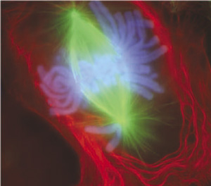
파일:Mitosis-fluorescent.jpg
Mitosis-fluorescent.jpg (300 × 264 픽셀, 파일 크기: 21 KB, MIME 종류: image/jpeg)
파일 역사
날짜/시간 링크를 클릭하면 해당 시간의 파일을 볼 수 있습니다.
| 날짜/시간 | 섬네일 | 크기 | 사용자 | 설명 | |
|---|---|---|---|---|---|
| 현재 | 2006년 1월 25일 (수) 05:25 |  | 300 × 264 (21 KB) | Mortadelo2005 | An image of a newt lung cell stained with flourescent dyes undergoing mitosis, specifically during early anaphase. According to NIH, "The scientists use newt lung cells in their studies because |
이 파일을 사용하는 문서
다음 문서 1개가 이 파일을 사용하고 있습니다:
이 파일을 사용하고 있는 모든 위키의 문서 목록
다음 위키에서 이 파일을 사용하고 있습니다:
- anp.wiki.x.io에서 이 파일을 사용하고 있는 문서 목록
- ast.wiki.x.io에서 이 파일을 사용하고 있는 문서 목록
- bn.wiki.x.io에서 이 파일을 사용하고 있는 문서 목록
- ca.wiki.x.io에서 이 파일을 사용하고 있는 문서 목록
- de.wiki.x.io에서 이 파일을 사용하고 있는 문서 목록
- de.wiktionary.org에서 이 파일을 사용하고 있는 문서 목록
- el.wiki.x.io에서 이 파일을 사용하고 있는 문서 목록
- en.wiki.x.io에서 이 파일을 사용하고 있는 문서 목록
- en.wikibooks.org에서 이 파일을 사용하고 있는 문서 목록
- eo.wiki.x.io에서 이 파일을 사용하고 있는 문서 목록
- es.wiki.x.io에서 이 파일을 사용하고 있는 문서 목록
- eu.wiki.x.io에서 이 파일을 사용하고 있는 문서 목록
- fa.wiki.x.io에서 이 파일을 사용하고 있는 문서 목록
- fr.wiki.x.io에서 이 파일을 사용하고 있는 문서 목록
- fr.wiktionary.org에서 이 파일을 사용하고 있는 문서 목록
- gl.wiki.x.io에서 이 파일을 사용하고 있는 문서 목록
- gu.wiki.x.io에서 이 파일을 사용하고 있는 문서 목록
- hi.wiki.x.io에서 이 파일을 사용하고 있는 문서 목록
- hu.wiki.x.io에서 이 파일을 사용하고 있는 문서 목록
- it.wiki.x.io에서 이 파일을 사용하고 있는 문서 목록
이 파일의 더 많은 사용 내역을 봅니다.


