동공 확대근
(동공확대근에서 넘어옴)
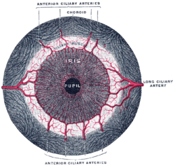
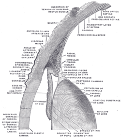
동공 확대근[2](iris dilator muscle (pupil dilator muscle, pupillary dilator, radial muscle of iris, radiating fibers, 동공 산대근)은 눈의 평활근[3]으로 홍채에서 방사형으로 움직이므로 확장기로 적합하다. 동공 확장기는 근상피 세포라고 하는 변형된 수축 세포의 바퀴살 모양 배열로 구성된다. 이 세포는 교감신경계에 의해 자극된다.[4] 자극을 받으면 세포가 수축하여 동공이 넓어지고 눈에 더 많은 빛이 들어오게 된다.
| Iris dilator muscle | |
|---|---|
 Iris, front view. (Muscle visible but not labeled.) | |
 The upper half of a sagittal section through the front of the eyeball. (Iris dilator muscle is NOT labeled and not to be confused with "Radiating fibers" labeled near center, which are part of the ciliary muscle.) | |
| 정보 | |
| 이는곳 | Outer margins of iris[1] |
| 닿는곳 | Inner margins of iris[1] |
| 신경 | Long ciliary nerves (sympathetics) |
| 작용 | Dilates pupil |
| 대항근 | Iris sphincter muscle |
| 식별자 | |
| 라틴어 | musculus dilatator pupillae |
| TA98 | A15.2.03.030 |
| TA2 | 6763 |
| FMA | 49158 |
섬모체근, 동공조임근 및 동공확대근은 때때로 내안근(intrinsic ocular muscle[5] 또는 intraocular muscle[6])이라고도 한다.
같이 보기
편집각주
편집- ↑ 가 나 Gest, Thomas R; Burkel, William E. (2000). “Anatomy Tables – Eye”. 《Medical Gross Anatomy》. University of Michigan Medical School. 2010년 5월 26일에 원본 문서에서 보존된 문서.
- ↑ 대한의협 의학용어 사전, 대한해부학회 의학용어 사전 https://www.kmle.co.kr/search.php?Search=Iris+dilator+muscle&EbookTerminology=YES&DictAll=YES&DictAbbreviationAll=YES&DictDefAll=YES&DictNownuri=YES&DictWordNet=YES
- ↑ Pilar, G; Nuñez, R; McLennan, I. S.; Meriney, S. D. (1987). “Muscarinic and nicotinic synaptic activation of the developing chicken iris”. 《The Journal of Neuroscience》 7 (12): 3813–3826. doi:10.1523/JNEUROSCI.07-12-03813.1987. PMC 6569112. PMID 2826718.
- ↑ Saladin, Kenneth (2012). 《Anatomy and Physiology》. McGraw-Hill. 616–617쪽.
- ↑ Barry D. Kels, Andrzej Grzybowski, Jane M. Grant-Kels. (March–April 2015). Human ocular anatomy. Clinics in Dermatology, Volume 33, Issue 2, Pages 140-146. https://www.sciencedirect.com/science/article/abs/pii/S0738081X1400234X
- ↑ Parker E. Ludwig; Sanah Aslam; Craig N. Czyz. (August 7, 2023.). Anatomy, Head and Neck: Eye Muscles. StatPearls Publishing; 2024 Jan-. https://www.ncbi.nlm.nih.gov/books/NBK470534/